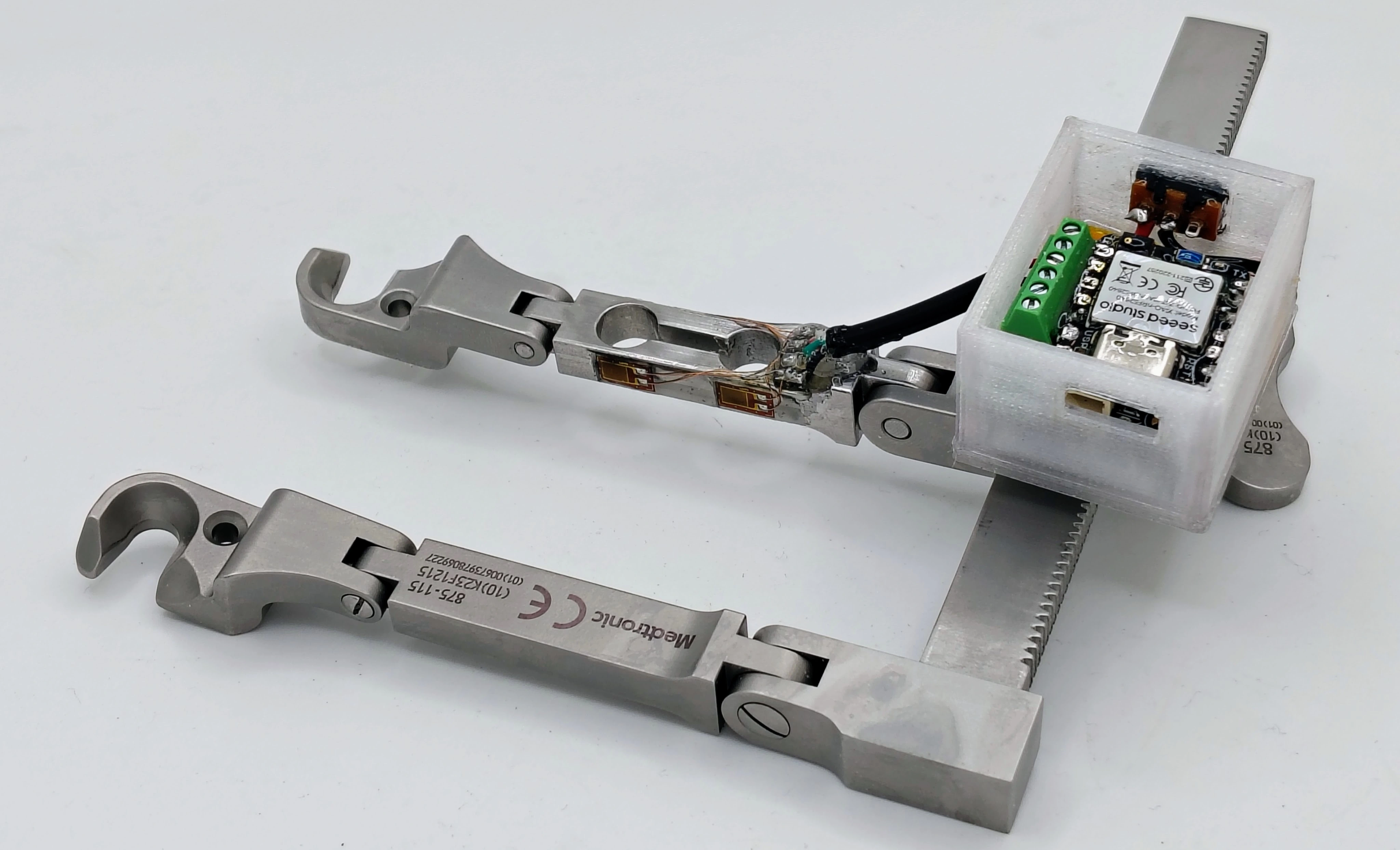
Tissue Pressure Monitoring Retractor
Project Overview
This project was completed in collaboration with Medtronic Spine R&D and focused on improving the Medtronic Trimline Retractor used during anterior cervical discectomy and fusion (ACDF) procedures.
The Trimline Retractor is a self-retaining surgical retractor designed to hold back the trachea, esophagus, muscles, and surrounding soft tissue, allowing surgeons to access the cervical spine. Once positioned, the device maintains retraction throughout the procedure while the surgeon performs the discectomy and fusion.
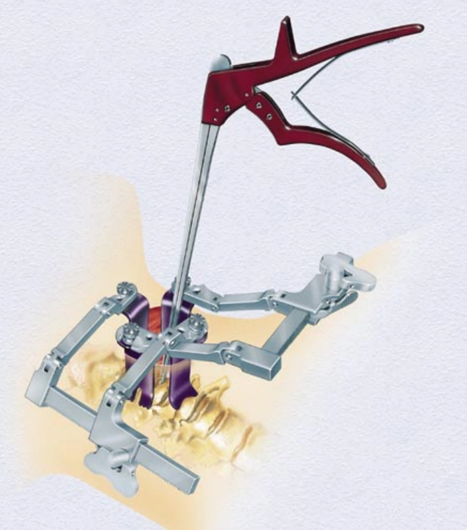
Source: Medtronic, TrimLine Anterior Cervical Discectomy & Fusion Instrument Set (2002)
Demonstration of the Medtronic Trimline Retractor during deployment.
One challenge with existing cervical retractors is that surgeons receive no quantitative feedback regarding the amount of force being applied to tissue or how long that tissue has remained under load. Excessive retraction force and prolonged loading have been associated with postoperative complications such as dysphagia (difficulty swallowing) and dysphonia (voice impairment), yet current systems rely entirely on surgeon experience and judgment.
This project explored two pressure-sensing modifications to the Trimline Retractor capable of providing real-time intraoperative feedback. The objective was to quantify tissue loading during surgery without altering the normal function, geometry, or workflow of the existing device.
Clinical Motivation
Anterior cervical spine procedures require prolonged soft-tissue retraction to expose the surgical site. While necessary for access, this retraction can place significant stress on surrounding anatomy.
Because current retractors provide no force feedback, surgeons cannot directly quantify tissue loading during a procedure. Developing a method to monitor pressure and time-under-load may help future studies identify relationships between retraction conditions and postoperative outcomes.
Design Requirements
The proposed system was required to:
- Maintain normal retractor functionality
- Provide real-time force feedback
- Remain compatible with existing surgical workflows
- Utilize sterilizable and biocompatible materials
- Avoid interference with the surgical field
- Operate safely in a clinical environment
Because the device would be used during surgery, preserving the geometry and usability of the existing retractor was considered a critical design constraint.
Design Concepts
Two sensing approaches were developed.
Concept 1: Instrumented Retractor Arm
The first concept integrated a strain-gauge load cell directly into the retractor rack. A structural component within the retractor assembly was replaced with a custom load cell designed to match the geometry of the original part.
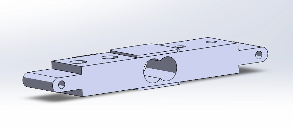
Load Cell Concept
Prototype Development
Before pursuing a CNC-machined stainless steel design, the load-cell concept was validated using a rapid FFF prototype printed in PETG. The prototype replicated the geometry of the proposed load cell and was instrumented with strain gauges to evaluate the sensing approach before committing to metal fabrication.
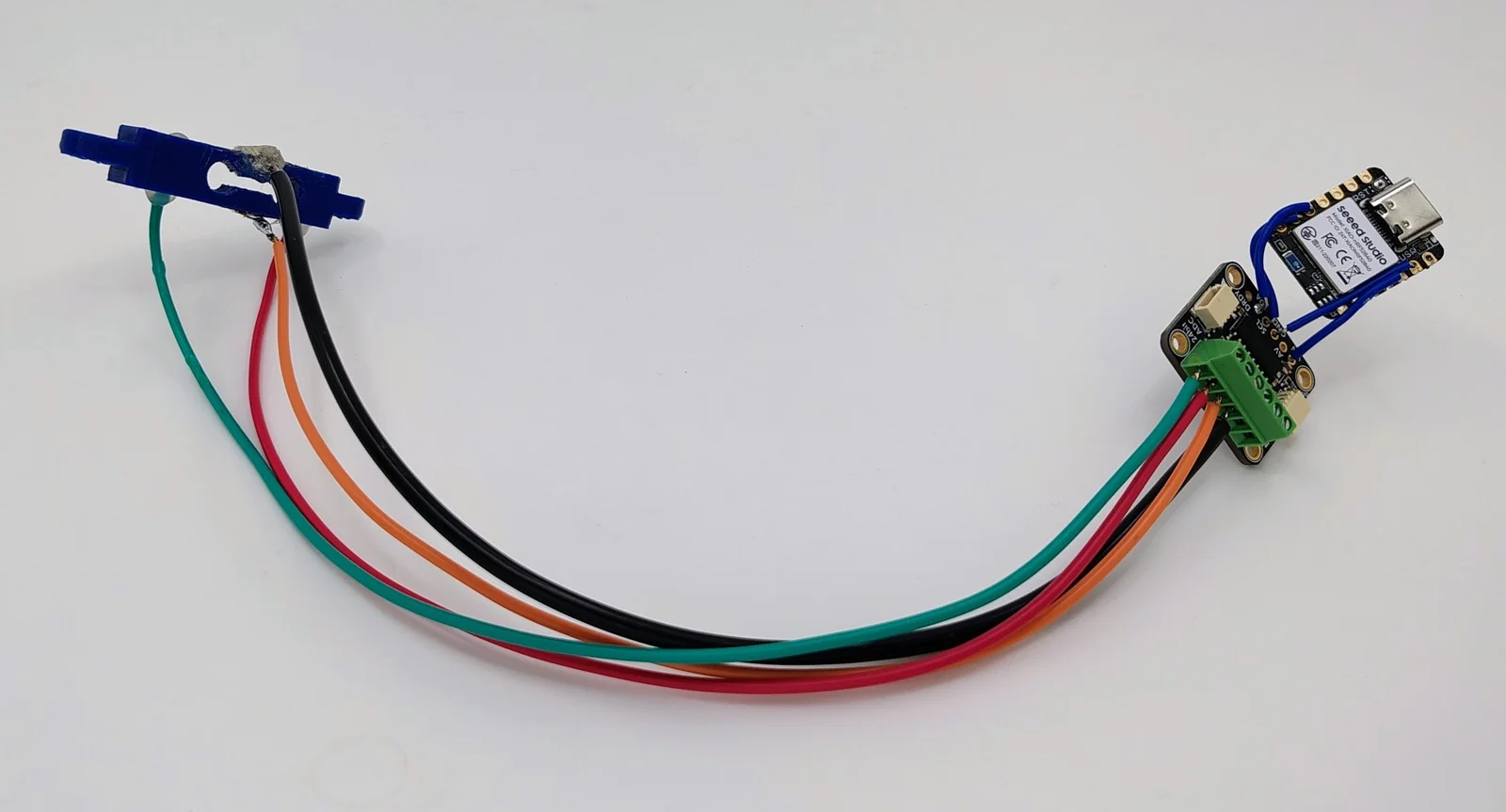
FFF-printed PETG prototype used for early validation testing.
The prototype demonstrated that measurable strain could be captured and converted into force measurements in real time. This early proof-of-concept reduced development risk and provided confidence that the sensing architecture would function as intended before moving to a CNC-machined stainless steel implementation.
As retraction force increased, the load cell experienced microscopic deformation that could be measured using bonded strain gauges. This approach provided a single quantitative measurement of the overall force applied through the retractor.
Concept 2: Pressure-Sensing Blade
The second concept incorporated an array of piezoelectric sensors directly into the retractor blade.
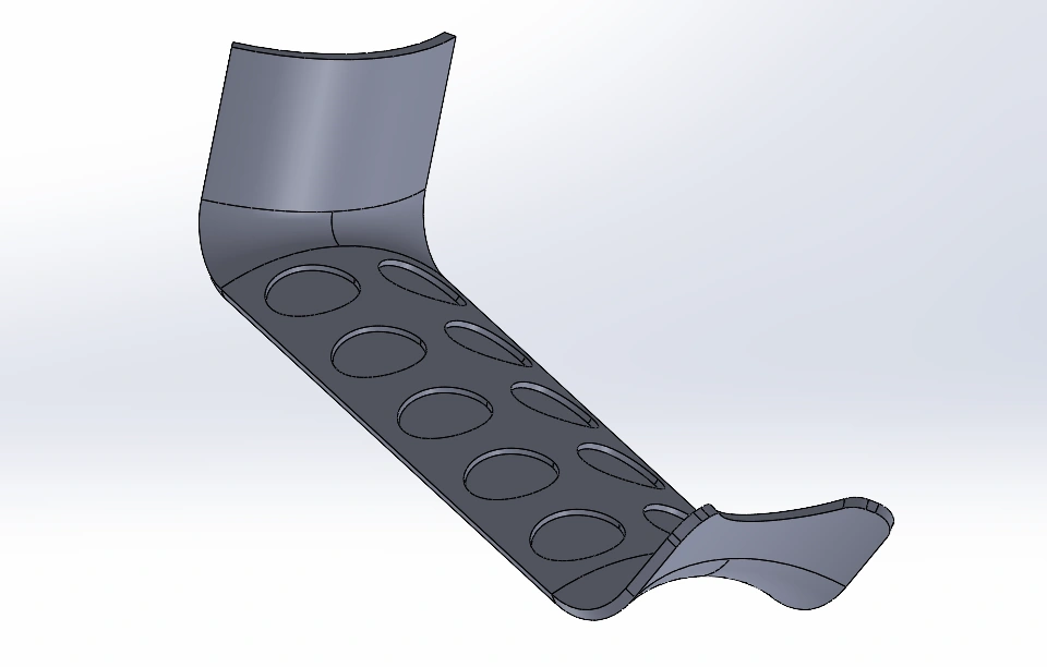
Retractor Blade Concept
Unlike the load-cell approach, this design provided spatial information about tissue loading. By arranging sensors across the blade surface, a pressure map could be generated to identify localized regions of high stress.
Electronics Architecture
Both concepts utilized a Silicon Labs EFR32BG29 wireless SoC microcontroller to acquire sensor data and transmit measurements to an external processing system.
For the load-cell concept, the signal pathway consisted of:
- Strain Gauges – Convert mechanical deformation of the load cell into small resistance changes.
- Bridge Amplifier / ADC – Amplifies the Wheatstone bridge output and converts the analog signal into a digital measurement.
- Microcontroller – Processes sensor data and transmits measurements wirelessly to an external computer.
The wireless architecture was selected to:
- Reduce cable clutter in the operating room
- Enable real-time monitoring
- Support future data logging and visualization capabilities
Sensor data would be processed on a connected computer and displayed through a surgeon-facing user interface.
Fabrication Strategy
Load Cell Assembly
The load-cell concept was designed around a CNC-machined 316L stainless steel structure.
The manufacturing process consisted of:
- CNC machining the load-cell geometry
- Stress-relief annealing
- Bonding foil strain gauges to the surface
- Wiring the gauges into a Wheatstone bridge circuit
- Signal amplification using an instrumentation amplifier
This approach leveraged established load-cell manufacturing techniques while maintaining compatibility with surgical sterilization requirements.
Pressure-Sensing Blade
The pressure-sensing blade was also designed to be fabricated from CNC-machined 316L stainless steel.
Piezoelectric sensors would be bonded directly to the blade surface in a grid pattern and connected to the microcontroller through dedicated analog channels.
The resulting design would generate a distributed pressure map rather than a single force measurement.
Proposed Testing
A staged testing approach was developed to evaluate the device.
Mechanical Validation
Initial testing would verify:
- Structural integrity
- Sensor calibration
- Electrical safety
Known loads would be applied to establish sensor accuracy and repeatability.
Tissue Testing
Following bench validation, the device would be evaluated using:
- Tissue phantoms
- Cadaveric models
- Porcine tissue models
These tests would assess the system’s ability to measure realistic tissue pressures while maintaining normal surgical operation.
Market Opportunity
Although ACDF retractors represent a specialized surgical market, they address a clinically significant problem that currently lacks a dedicated sensing solution.
The proposed platform could potentially be expanded beyond cervical spine surgery into other procedures requiring prolonged tissue retraction, creating opportunities within the broader surgical instrumentation market.
Key Takeaways
- Current ACDF retractors provide no feedback regarding tissue loading.
- Real-time pressure monitoring may help reduce preventable soft-tissue injury.
- Two complementary sensing approaches were developed: load-cell measurement and pressure mapping.
- The proposed designs preserve existing surgical workflows while adding quantitative feedback.
- Future work would focus on prototype fabrication, validation, and cadaveric testing.
Conclusion
This project explored two approaches for measuring tissue pressure during anterior cervical spine surgery. Both concepts sought to provide surgeons with real-time information regarding retraction forces while preserving the functionality of existing retractors.
Although the designs remain at the conceptual stage, they establish a foundation for future prototype development and testing. By introducing objective measurements into a procedure that currently relies entirely on surgeon judgment, pressure-sensing retractors have the potential to improve surgical decision-making and reduce postoperative complications.
Additional Documentation
This page summarizes the primary design concepts and outcomes of the project. A more detailed discussion of the clinical background, design process, testing strategy, regulatory considerations, and commercialization pathway can be found in the full technical report.